A0–1
What Is Light?
Discover the fundamental nature of light and why it is the most important tool in optical microscopy. Introduces light as an electromagnetic wave and explores wavelength, phase, and amplitude.
Microscopy academy · Eric Muzipov, 2026
A structured path from the nature of light to advanced optical techniques and scanning electron microscopy.
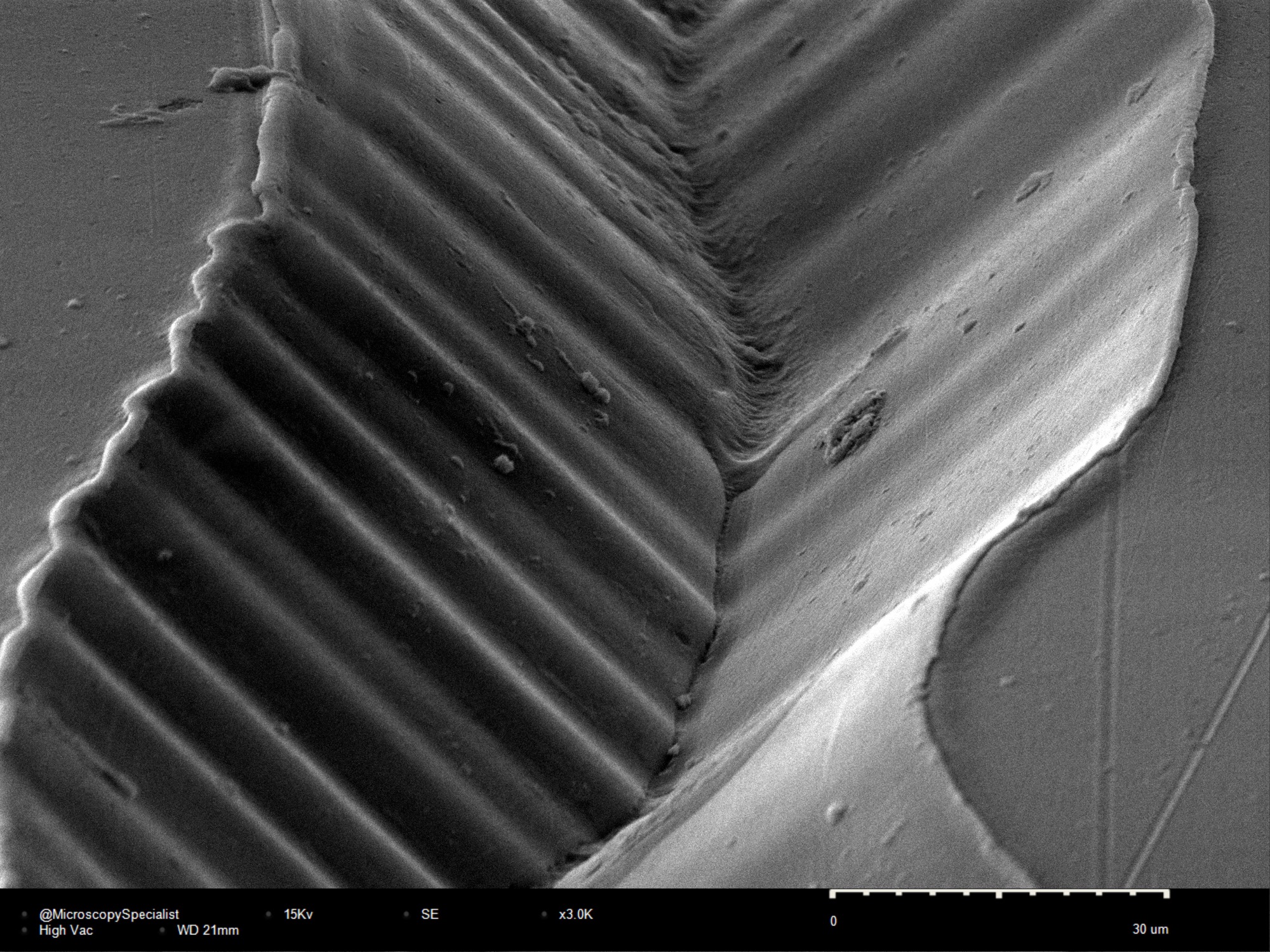
Original materials, animations, graphics, and practical examples developed from real work at the instrument.
A0–1
Discover the fundamental nature of light and why it is the most important tool in optical microscopy. Introduces light as an electromagnetic wave and explores wavelength, phase, and amplitude.
A0–2
Learn the optical phenomena that govern image formation inside a microscope — essential principles for interpreting image quality, contrast, and resolution.
A0–3
Explore illumination from traditional halogen lamps to modern LEDs, HBO, HXP, and lasers, and learn how the light source affects image quality and possible applications.
A0–4
Take a closer look at the components that guide and manipulate light: lenses, apertures, prisms, filters, and other key optical elements.
A0–5
Understand the difference between making an image larger and revealing more detail, including the factors that determine resolution and the practical limits of optical imaging.
A0–6
Discover the imperfections that degrade microscope images, the most common aberrations, and how modern objectives are designed to minimize them.
A1–1
Learn how objectives determine image quality, magnification, resolution, and contrast in microscopy.
A1–2
Understand how immersion media improve optical performance and increase resolution.
A1–3
Explore the most common microscope types and their typical applications.
A1–4
Get familiar with the main components and learn the fundamentals of operating a light microscope.
A1–5
Learn the principles of brightfield microscopy and how to set up Köhler illumination for the best image quality.
A2–1
Learn how contrast techniques use condensers and optical inserts to reveal features that remain invisible in brightfield microscopy.
A2–2
Discover how darkfield illumination enhances transparent samples by imaging only scattered light.
A2–3
Learn how phase contrast converts invisible phase differences into visible image contrast without staining.
A2–4
Explore how polarized light reveals crystal structure, stress, and anisotropic properties in materials.
A2–5
Understand how DIC, or Nomarski contrast, creates high-contrast images with a three-dimensional appearance by detecting optical path differences.
A2–6
Learn the principles of reflected light microscopy and its applications in materials science, metallurgy, and surface inspection.
B1–1
Learn how fluorescent molecules make it possible to visualize specific structures with exceptional sensitivity and contrast.
B1–2
Explore microscope cameras, sensors, and the imaging parameters that influence image quality and acquisition speed.
B1–3
Understand how confocal microscopy removes out-of-focus light, improves contrast, and enables optical sectioning.
B1–4
Take an in-depth look at stereo microscope optics, configurations, and practical applications.
B1–5
Learn how to select and combine microscope components into an optimized system for a specific application.
B2–1
Explore advanced optical methods that improve resolution and contrast beyond conventional microscopy.
B2–2
An introduction to electron microscopy, its underlying principles, major components, and capabilities.
B2–3
Learn how imaging parameters influence image quality and how to optimize SEM acquisition settings.
B2–4
Discover how low-vacuum operation enables imaging of non-conductive and challenging samples with minimal preparation.
B2–5
Compare FEG and tungsten-filament SEM, explore specialized imaging modes, and see how SEM and STEM are applied across different fields.
Enrollment
Course details and enrollment updates are available on the Microscopy Academy website.
Visit the Academy